Joint Fluid Analysis
Test Overview
Joint fluid analysis is a test to look at joint fluid under a microscope for problems such as infection, gout, pseudogout, inflammation, or bleeding. The test can help find the cause of joint pain or swelling.
Normally, only a small amount of joint fluid is found in a joint. Joint fluid acts as a lubricant for the joint and cushions joint structures. If you have a joint problem, you may have more fluid in your joint and your joint may become swollen, stiff, and painful.
A sample of joint fluid can be taken from any joint in your body. The joint fluid is then analyzed in a lab to look for inflammation, infection, gout, pseudogout, or bleeding.
Why It Is Done
This test is done to find inflammation, infection, gout, or pseudogout. Removing some of the joint fluid may also relieve pain caused by the buildup of fluid in your joint.
How To Prepare
- In general, you don't need to prepare before having this test. Your doctor may give you some specific instructions.
- If you take a medicine that prevents blood clots, your doctor may tell you to stop taking it before your test. Or your doctor may tell you to keep taking it. (These medicines include aspirin and other blood thinners.) Make sure that you understand exactly what your doctor wants you to do.
How It Is Done
Joint fluid analysis can be done in your doctor's office, clinic, operating room, or emergency room. Depending on which joint will be examined, you may be asked to undress and put on a hospital gown. You will sit or lie down on an examining table.
Your doctor will examine the joint to find out where the needle should be inserted. Your doctor may use ultrasound to guide the needle placement. The skin over the joint area will be cleaned with antiseptic solution. A local anesthetic is often injected into the skin over the joint. For young children, a sedative may also be given.
A long, thin needle is slowly inserted in the joint area. A syringe attached to the needle is used to remove a sample of joint fluid. Samples of the fluid may be put in special tubes or containers and sent to the lab. A cortisone shot may be given into the joint before the needle is removed. It can help keep fluid from building up again.
A tight (pressure) bandage will be placed over the site to reduce swelling and bruising. An elastic bandage may also be wrapped around your joint, such as your knee, to reduce swelling.
How long the test takes
- The test will take about 20 minutes.
How It Feels
You will feel a prick or sting when the anesthetic is given. You may feel tingling, pressure, pain, or fullness in your joint as the fluid is removed.
Risks
There is very little chance of having a problem after a joint fluid analysis. Infection, bleeding, or damage to the tendon, nerve, or joint is rare.
Sometimes the doctor may not be able to draw any fluid out. The joint space may be too small, or you may have scar tissue in the joint space. Or there may not be any fluid in the joint.
Results
The results of a joint fluid analysis are usually ready the same day. The results from a culture usually take a few days.
| ​ | Normal | Abnormal |
|---|---|---|
Color and clarity | Clear to light yellow | Red (bloody) or milky white (cloudy) |
Blood cell count | No large numbers of red or white blood cells | Large numbers of red or white blood cells |
Crystals (seen under a special microscope with polarized light) | Not present | Present |
Gram stain and culture | No bacteria are seen, and no organisms grow in the culture. | Bacteria are seen, or organisms grow in the culture. |
Abnormal values
- Color and clarity.
Slightly cloudy fluid may be caused by inflammation, gout, or pseudogout. A deep, dark red color may be caused by bleeding in the joint. Milky white may be caused by infection or inflammation or a condition such as gout.
- Blood cell count.
Large numbers of red blood cells may be caused by bleeding in the joint from injury, inflammation, or abnormal clotting of the blood. Large numbers of white blood cells may be caused by gout, pseudogout, other types of arthritis (such as rheumatoid arthritis), psoriatic arthritis, injury, or infection.
- Presence of crystals.
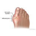
Uric acid crystals in the joint mean you have gout. Calcium pyrophosphate crystals mean you have pseudogout.
- Gram stain and culture.
Bacteria in the joint fluid that are causing an infection may be seen under a microscope after being colored with a Gram stain (a special dye). Joint fluid added to a substance that promotes the growth of germs (such as bacteria or a fungus) may show an infection. This is called a culture.
Credits
Current as of: April 30, 2024
Author: Ignite Healthwise, LLC Staff
Clinical Review Board
All Ignite Healthwise, LLC education is reviewed by a team that includes physicians, nurses, advanced practitioners, registered dieticians, and other healthcare professionals.
Current as of: April 30, 2024
Author: Ignite Healthwise, LLC Staff
Clinical Review Board
All Ignite Healthwise, LLC education is reviewed by a team that includes physicians, nurses, advanced practitioners, registered dieticians, and other healthcare professionals.


